July 21, 2022
Infected for Science
Viruses play an important role in experimental biology acting as sneaky couriers for genetic material
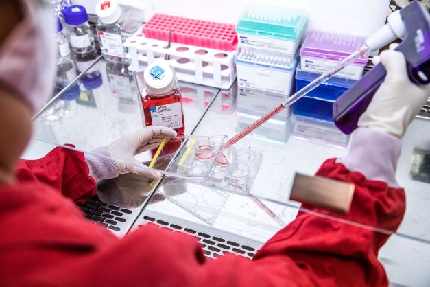
A team at the Institute of Science and Technology Austria (ISTA) is cooking up viruses in a lab – not for evil plans of world domination, but for science! In their viral kitchen, the Virus Services team produces many different kinds of viruses for the Institute’s scientists who use them to smuggle genetic material into cells to track their growth, movement, and activity.
Viruses – especially the coronavirus – have been in the news for some time now. While we usually associate viruses with diseases, scientists use them as tools in their research. Viruses are amazing little machines that can do things nobody else can do. At the Institute of Science and Technology Austria (ISTA), the Virus Services team provides scientists with the viral help they need to do their work.
There are myriads of different viruses, from the ones causing the common cold to HIV or the well-known coronavirus to many more that do not cause diseases in humans at all. While it is disputed among scientists whether viruses are technically alive or just molecular machines, it is clear that they are very efficient in infecting host cells and making them replicate the virus particles. The size of the particles range from a few dozen nanometers – a millionth of a millimeter – to several hundred nanometers. Most are smaller than the shortest wavelength of visible light (violet at about 380 nanometers) and yet they are enormously successful all around the globe. Their molecular machinery of replication, honed by millions of years of evolution, is what scientists make use of when employing viruses as their tiny helpers in the lab.

Building a Virus
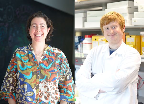
“Simply put, a virus particle consists of an outer protein shell, sometimes covered in an additional envelope made of fats, and the viral genome that encodes how to make more of it,” Flávia Leite, virus expert at ISTA, explains. “In the Virus Services lab, we use modified viruses, so they are no longer dangerous. Instead of their original genetic material, they carry some desired gene into the cells they infect. There, it can also get incorporated into the genetic code of an infected cell.” For unmodified viruses, the infected cells would then create more of the virus to spread it to other cells. In Flávia’s viruses, the genome encodes proteins that the researchers are interested in. This way, the virus cannot reproduce and spread any further making it safe to handle in experiments.
But how do you put this genetic material into the virus in the first place? Mark Smyth, another expert in the Virus Services team, explains, “In order to create, for example, lentivirus particles, we use three different building blocks called plasmids. These are short, circular strands of DNA. One contains the genetic material which the researchers want to put into the cell, one encodes the virus’s shell, and one encodes other proteins needed to replicate viral genetic material and pack it into the viral particles. Only when all three plasmids come together in a host cell, they take over and the cell produces the virus particles. We can even get plasmids from researchers outside ISTA and incorporate them into our viruses. It’s like cooking, but with viruses.” Doing it that way also has the advantage of never having the original and dangerous virus assembled in its full form. By dealing only with its components, this method is very safe.
Marking Cells
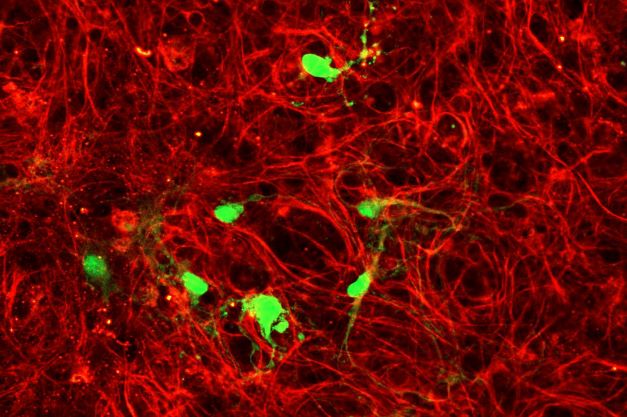
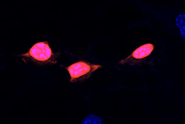
“We use several different kinds of viruses,” Smyth explains. “For example, researchers use adeno-associated viruses to make the targeted cells produce green fluorescent protein (GFP).” GFP can be seen in the microscope by looking for the green glow which it emits under ultraviolet light. With AAV viruses, GFP is normally used to check that the virus successfully infected the cell and consequently that the protein of interest was successfully introduced or to mark specific cells. GFP is not the only fluorescent protein. There are also proteins which glow blue, red or yellow under ultraviolet light.
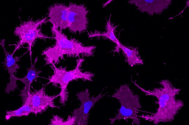
“The virus acts as a cellular tool,” Leite adds. “Using them, you can turn off a gene to understand what role it plays in the cell or we can even make certain genes turn on and off only using flashes of light.” Adeno-associated viruses are especially useful for neuroscientists because their different types target different kinds of brain cells. For example, one type preferentially infects cells in the hippocampus and another targets microglia cells that are part of the brain’s immune system.
Another type of virus the Virus Services team produces is called lentivirus which includes the human immunodeficiency virus (HIV). “We use this virus because it can introduce DNA into both dividing and non-dividing cells – something other viruses cannot do,” Leite says. “It also has the special ability to make the DNA it carries a permanent part of the infected cell’s genome. This way, the target DNA gets passed on to the cell’s descendants and we can create whole modified cell lines.” Again, scientists often use lentiviruses to make the cells produce green fluorescent protein together with another target protein. For example at ISTA, the group led by Sandra Siegert uses them to track microglia immune cells in the brain and the group around Michael Sixt used lentivirus to track immune cells moving through dense tissue.
The third kind of virus the Virus Services team produces is the rabies virus. While being very dangerous in its natural form, in the lab, it can help neuroscientists to map connections in the brain. Smyth explains, “Rabies virus is highly adapted to attack the nervous system and when applied correctly it infects neurons in the direction opposite to the information flow in the brain.” So, the virus moves from the infected neuron to the other neurons that send information to it. The information pathway becomes visible by making the neurons produce a fluorescent protein that the researchers can see under the microscope. Neuroscientists use this remarkable behavior to find individual signal pathways in the brain.
Established just in 2019, the Virus Services team has become an important partner for the experimental biologists at ISTA. “Having such a facility in-house is not that common and in the Vienna area, we are the only one,” Leite remarks. “Usually, such work is outsourced to companies, but having it done on the ISTA campus allows us to work very closely with the scientists. We can advise the researchers which virus to use and create custom solutions, while we also get valuable feedback from them that allows us to expand our offer.” Flávia Leite has a long career working with viruses working as a Postdoc at The Francis Crick Institute before joining ISTA in 2019. Mark Smyth is finishing his PhD on the evolution of viruses at the Research Center for Molecular Medicine of the Austrian Academy of Sciences (CeMM) and he joined ISTA in 2021. They are supported by Rexhina Sinani working as a virus technician.
Animal welfare
In order to better understand fundamental processes, for example, in the fields of neuroscience, immunology, or genetics, the use of animals in research is indispensable. No other methods, such as in silico models, can serve as alternative. The animals are raised, kept, and treated according to the strict regulations of Austrian law. All animal procedures are approved by the Federal Ministry of Education, Science and Research.
